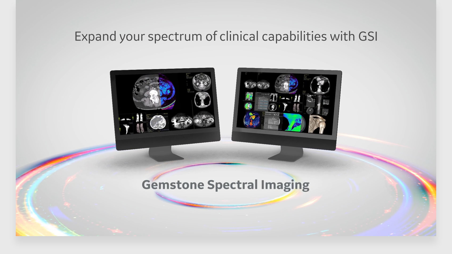
gsi viewer

Aids in confident diagnosis with easier workflow

Expand your spectrum of clinical capabilities with spectral imaging

Leverage the power of Spectral Imaging with 3D advanced visualization capabilities

  • GSI Gout Protocol

    Fully automated workflow for material decomposition and color overlay to assist in gout characterization:

    • Automated generation of 2D and 3D gout overlay
    • Automated export of 2D and 3D gout overlay images to the patient browser
  • GSI Pulmonary Perfusion²

    Dedicated one-click protocol protocol to assist in identification of relative hypo-perfused territories:

    • Automatic relative hypo-perfused region display
    • Automated export of 2D and 3D gout overlay images to the patient browser
    • Volumes reported for total lung and relative hypo-perfusion volumes
    • Succinctly communicate volumes and relevant pictures to referring physicians
  • GSI Fat

    Dedicated GSI fat capability to quantify fat in the liver:

    • Evaluate hepatic steatosis with liver fat percentage measurements
    • GSI with material separation provides ability to measure approximate liver fat percentages
    • Liver fat measurements compatible with non contrast¹ and multi-phase contrast acquisitions

Supporting Materials

  1. GSI Liver Fat ROI’s can be done on non-contrast or any phase of contrast enhancement.
  2. GSI Pulmonary Perfusion protocol is an optional add-on protocol to Thoracic VCAR application.

JB01114AU